
If you or your loved one worked as a tradesperson prior to 1985 and were diagnosed with lung cancer within the last 3 years you may be entitled to substantial financial settlement. Contact us today.
Lung Cancer Victims
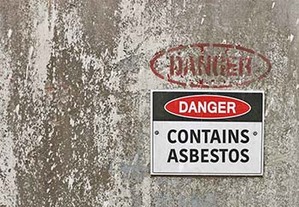
Lung Cancer has been linked to asbestos, a toxic mineral often unknowingly handled by people who worked in a wide variety of trades. Asbestos is a mineral fiber that might be found in construction materials, insulation, paint and automobiles, and can increase your risk of developing life-threatening diseases.
Some of the most common industries that require employees to handle asbestos are:
- Commercial construction
- Shipyards
- Chemical plants
- Auto repair
- Military service
- Power plants
- Oil refineries
According to the EPA, three major health effects of handling asbestos are:
- Lung cancer
- Mesothelioma, or cancer of the linings of the heart, lung, chest and abdomen
- Asbestosis, a non-cancer lung disease

- Automotive Industry
- Auto mechanic
- Boiler operator
- Construction
- Chemical plants
- Electrician
- Firefighter
- Machinist
- Maintenance worker
- Metal works
- Navy or other Military service
- Oil refineries
- Pipe-fitter
- Pipe-layer
- Plasterer
- Plumber
- Power plants
- Shipyards
- Tradesmen
- Welder
Some of the most common jobs that require employees to handle asbestos are:
Veterans Exposed to Asbestos: You Have Important Legal Rights
Every year on November 11, we remember all the sacrifices our veterans have made for us. From the 1930s through the 1970s, asbestos products were heavily used in the military for its heat resistant and insulating properties.
Non-Small Cell Lung Cancer, Explained
The most common type of lung cancer is non-small cell, making up 80-85% of lung cancer.
This cancer is categorized based on the size of the original tumor and where the cancer has spread.
Lung cancer patients may be able to improve survival and quality of life with a web application called Moovcare.
This web app was revealed at the annual American Society of Clinical Oncology meeting.
"The Law Firm of Pintas & Mullins has been very helpful... with the representation of my daughter’s case. Emily specifically has been very professional, informative, straightforward, compassionate, extremely courteous, helpful, and patient. If you are in need of good legal representation, I would highly recommend them."
T S

Kimberly
"Sheila really helped out getting my case together listened to everything took her time making sure I was comfortable informed me about the process. It's really great that they are right behind you helping you.. Thank you so much Sheila for your kindness and professionalism. I do feel like my claim is in the best hands."
"My experience was totally and genuinely pleasant. I spoke with a young lady named Ty... She was very patient and she also happened to give me optimism whether she knew it or not. I will gladly recommend this firm to anyone who not only needs a lawyer that's going to fight... but also one who will be empathetic"
Jamison
Complete the Form Above
Free Case Review
Get Justice
Time to file a claim is limited. Fill out the form above to request your free case review.
You will be contacted for your free case review
No fees are charged unless you are awarded compensation
In the last 12 months, we have recovered more than $250 million in settlements for our clients.


We are here to ensure that your voice is heard. If you or a loved one has developed lung cancer, we want to hear about it. Contact us today by submitting the form above, and we will review your case for free.

Hiring an attorney is a serious matter and should not be based solely upon advertising. Past performance is no guarantee of future results. In the absence of recovery, the client is NOT liable for attorney fees. Pintas & Mullins Law Firm PR, LLC is a law firm located at 250 Ave. Luis Muñoz Rivera, Suite 300, San Juan, PR 00918. William Pintas and Laura Mullins are licensed in Illinois and will maintain professional and financial responsibility on accepted cases even when they may partner with firms in other states where licensing may be required. Not available in all states.